
If you want to understand how a plant works, it helps to be able to see cells and tissues. That’s easy in a biology lab with microscopes and prepared slides.
Once you leave that world, though, it becomes more difficult to see the structures beneath things.
Wikipedia articles tend to be well-illustrated, with pictures of macroscopic plant structures—the kind of thing you can photograph with your average camera—but high quality photomicrographs are much rarer. It’s even more difficult to find images that can be freely reused in ways that help anyone learn.
Leslie Zeman’s Cellular Anatomy class class at the University of Washington helped tackle that problem. Student editors in that class created and uploaded images of the anatomical structures of plants.
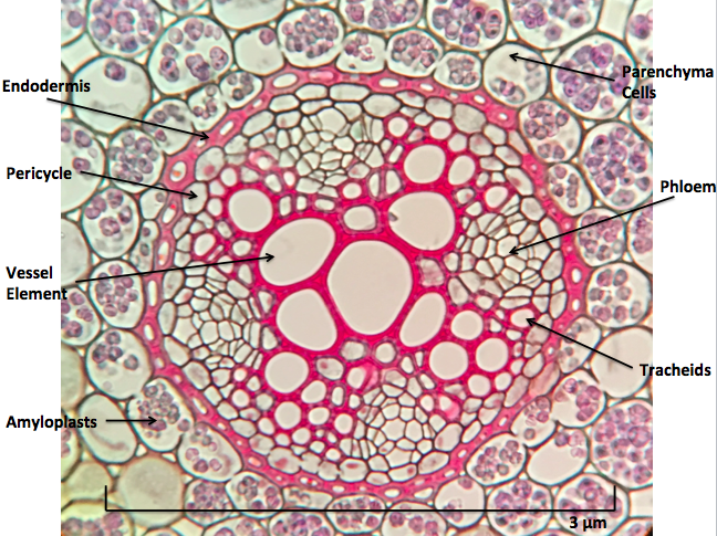
Cross sections of Ranunculus roots, like the one above, are commonly used to teach plant anatomy. This image was uploaded by students in that course.
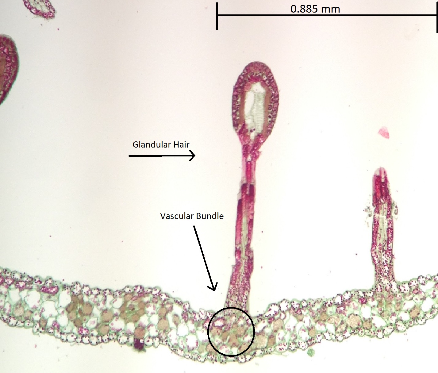
Sundews are carnivorous plants that trap insects using glandular hairs. The hairs are big enough to see with the naked eye, but a photomicrograph like the one above, uploaded by students in the class, give a sense of things at the cellular level.
From my own experience as an instructor, I know how challenging it can be to get students to engage with microscopy. Often, students are only motivated to look for what they’re “supposed to see,” and spend their time trying to match the slide to what’s in their textbook.
When students actually delve into the universe at the other end of the microscope, that’s when the magic happens. When you see the work that students in Leslie Zeman’s class have produced, you know you’re looking at the handiwork of enthusiastic explorers.
Wiki Ed’s tools make it easy to turn a good class assignment into a project with wider, more lasting implications. By adding their images to Wikimedia Commons, students in the class have done something that reaches outward from the microscope, out into the world. They’ve expanded the pool of freely reusable plant anatomy images. They’ve made images available for others to use and learn from, to create new teaching resources for all.
The Wikipedia Year of Science is all about making an individual student’s work more visible than it could ever be. When your students connect their learning to Wikipedia, they’re a part of something bigger than their classroom.
Want to see what your class can do? See how you can get started right away. Or send us an email: contact@wikiedu.org.
Top Photo: Vascular cambium of helianthus stem by Katrina Burkhardt – Own work, CC BY-SA 4.0.
Nicely done. Carol and Larry Peterson produced an excellent atlas of Plant Antomy using sections cut with razor blades and stained with various dyes to enhance certain types of cell walls, organelles, and so on. Both approaches, prepared slides of wax sections and hand sections of fresh material, make plant structure easier to understand and appreciate.